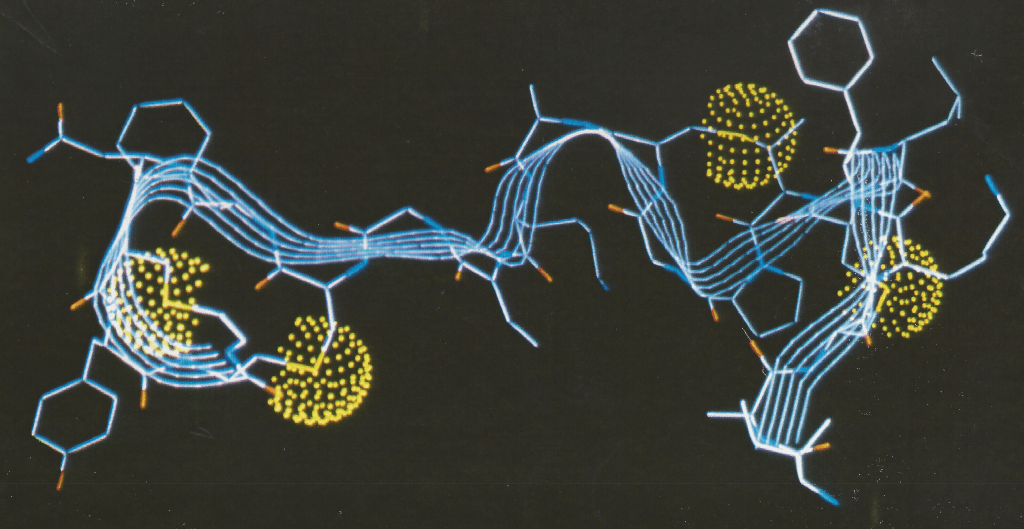
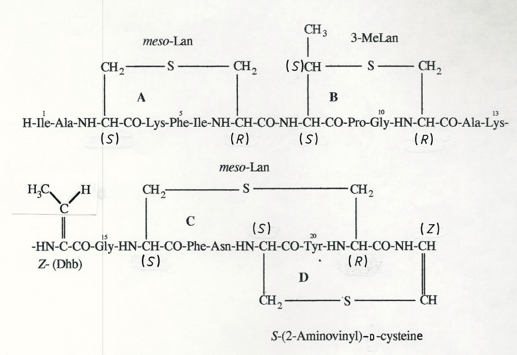
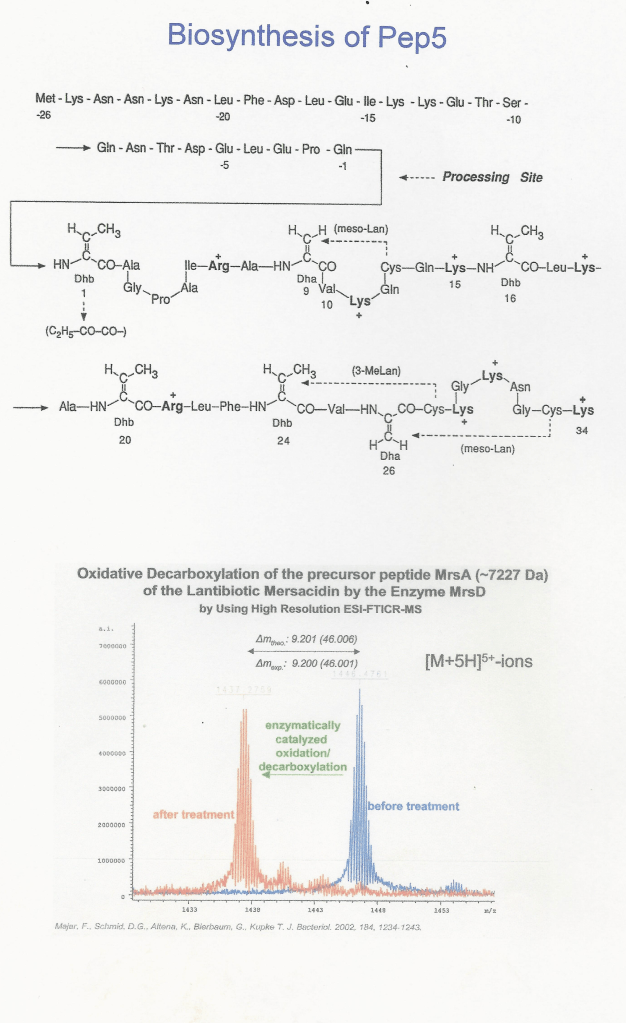
Microcins and lantibiotics are families of polypeptides, synthesized via precursor-proteins and posttranslational modifications at serine, threonine and cysteine residues. Thereby side chains and backbone are transformed to give sulphide rings, 2,3-didehydroamino acids, D-amino acids, oxazole and thiazol rings. 3D-structures of the resulting conformationally restricted polypeptides have been elucidated. In cooporation with microbiologists (F.Götz and H.-G. Sahl), their biosynthesis, the sequences of prepeptide intermediates, sequential analogues and importantly the novel peptide modifying enzymes were elucidated. Thus the molecular basis of a novel protein engineering with defined construction of unusually modified peptides was established.

N. Schnell, K-D- Entian, U. Schneider, F. Götz, H. Zähner, R. Kellner, and G. Jung
(1988). Prepeptide Sequence of Epidermin, a Ribosomally Synthesized Polypeptide Antibiotic Containing four Sulphide-Rings, Nature 333,276-278.
R. Kellner, G. Jung, M. Josten, C. Kaletta, K-D. Entian, and H.-G. Sahl (1989). Pep5:Structure Elucidation of a Large Lantibiotic, Angew. Chern. 101, 618-621; Angew. Chern. Int. Ed. Engl. 28, 616-619.
G. Jung (1991). Lantibiotics – Ribosomally Synthesized Biological Active Polypeptides containing Sulfide Bridges and α, β-Didehydroamino Acids, Angew. Chern. 103, 1067-1084; Angew. Chern. Int. Ed. Engl. 30, 1051-1068.
N. Zimmermann, S. Freund, A. Fredenhagen, and G. Jung (1993). Solution Structures of the Lantibiotics Duramycin B and C, Eur. J. Biochern. 216, 419-428.
G. Jung and H.-G. Sahl (1991) (eds.). Nisin und Novel Lantibiotics, 490 pp., Escorn, Leiden.
A. Bayer, S. Freund, G. Nicholson und G. Jung (1993). Posttranslational Backbone Modifications in the Ribosomal Biosynthesis of the Glycine-Rich Antibiotic Microcin B17, Angew. Chern. 105, 1410-1413; Angew. Chern. Int. Ed. Engl.32, 1336-1339.
M. Skaugen, J. Nissen-Meyer, G. Jung, S. Stevanovic, K.Sletten, C.I Mortvedt Abild-
gaard, and I.F. Nes (1994). In vivo Conversion of L-Serine to D-Alanine in a Ribosomally Synthesized Polypeptide, J. Biol. Chern. 269, 27183-27185.
A. Bayer, S. Freund, and G. Jung (1995). Post-translational Heterocyclic Backbone Modifications in the 43-Peptide Antibiotic Microcin B17, Structure Elucidation and NMR Study of a 13C, 15N-Iabelled Gyrase Inhibitor, Eur. J. Biochem.234, 414-426.
T. Kupke, C. Kempter, G. Jung, and F. Götz (1995). Oxidative Decarboxylation of Peptides Catalyzed by Flavoprotein Epi D: Determination of Substrate Specificity Using Peptide Libraries and Neutral Loss Mass Spectrometry, J. Biol. Chem. 270, 11282-11289.
B.Ottenwälder, T. Kupke, S. Brecht, V. Gnau, J. Metzger, G. Jung, and F. Götz (1995).
Isolation and Characterization of Genetically Engineered Gallidermin and Epi-
dermin Analogs, Appl. Environ. Microbiol. 61, 3894–3903.
N. Zimmermann, J.W. Metzger, and G. Jung (1995). The Tetracyclic Lantibiotic Actagardine, 1NMR and 13C-NMR Assignments and Revised Primary Structure, Eur. J. Biochem., 228, 786-797.
G. Videnov, D. Kaiser, C. Kempter, M. Brooks, and G. Jung (1996). Synthesis of the DNA Gyrase Inhibitor Microcin B17, a 43-Peptide Antibiotic with Eight Heteroaromatic Rings in the Backbone, Angew. Chem., 108, 1607-1609; Angew. Chem. Int. Ed. Engl. 35, 1506-1508.
G. Bierbaum, C. Szekat, M. Josten, C. Heidrich, C. Kempter, G. Jung, and H.-G. Sahl
(1996). Engineering of a Novel Thioether Bridge and Role of Modified Residues in the
Lantibiotic Pep5, Appl. Environm. Microbiol., 62, 385-392.
F.J.M. van de Ven and G. Jung (1996). Structures of Lantibiotics Studied by NMR , Antonie van Leeuwenhoek 1. Microbiol., 69, 99-107.
D.G. Schmid, F. Majer, T. Kupke, and G. Jung (2002). Electrospray Ionization Fourier Transform Ion Cyclotron Resonance Mass Spectrometry to Reveal the Substrate Specificity of the Peptidyl-cysteine Decarboxylase EpiD, Rap. Comm. Mass Spectr. 16, 1779-1784.
G. Jung (2006). Enzyme-Catalyzed Sulfide Ring Formation in Lantibiotics, Angew. Chem. 118, 2-5; Angew. Chem. Int. Ed. 45, 5919-5921.
Biosynthesis of McB17

Solution Structure of Gallidermin and Sequence of Epidermin